Special eye tests mean extra special eye care!
ZEISS / Humphrey Visual Field Testing
A visual field test measures peripheral or side vision while detecting the loss or alteration of nerves in the eye and the function of retinal tissue. The field test is essential in identifying peripheral vision loss and provides a detailed map of the extent and depth of loss while diagnosing diseases such as glaucoma or macular degeneration. In addition, it is useful when a patient has unexplained visual disturbances or vision loss and can quickly and accurately detect neurologic disorders such as tumors, strokes and aneurysms.
Retinal Digital Imaging – Fundus Photography
Fundus photography documents the neurosensory tissue in our eyes that translates the optical images we see into the electrical impulses our brain understands. The retina can be photographed directly as the pupil is used as both an entrance and exit for the Fundus camera’s illuminating and imaging light rays. Ophthalmologists use these retinal photographs to follow, diagnose and treat eye diseases.
Fundus photography can be performed with colored filters, or with specialized dyes including fluorescein and indocyanine green.
Genetic Testing
With the evolution of genetic testing comes the ability to more precisely diagnose your patients for inherited ocular diseases.
Macular Degeneration Testing
Macular Degeneration is a progressive and degenerative disease affecting the central part of the retina, the macula. It is the leading cause of irreversible vision loss in the United States.
Age-Related Macular Degeneration (AMD) affects 11 million Americans. 70% of the cause of AMD is due to genetics. This puts 20 million Americans at risk of AMD due to family history. Our hereditary AMD genetic tests can help you better understand your risk of macular degeneration and plan effective treatment.
AMDiGuard DNA Risk Test
The AMDiGuard Risk Test is designed to use DNA through a simple swab combined with patients’ basic health factors and provide a risk level of aged-related macular degeneration based on our scientific analysis of both factors.
AMDiGuard DNA Progression Test
The AMDiGuard Progression Test is designed to learn more about the risk of advanced AMD for patients who have early to intermediate AMD.
Retinal Disease Testing
With the evolution of genetic testing comes the ability to more precisely diagnose your patients for inherited retinal diseases.
More answers from testing or retesting may uncover more possibilities for active clinical trials, emerging treatments, and even identifying underlying conditions beyond vision issues.
Genetic testing may give patients more ANSWERS:
- Affirm the genetic cause of vision loss or impairment
- Navigate new insights into how vision may change over time
- See if you are eligible for clinical trials and emerging treatments
- Weigh the potential risks of other family members
- Education about support groups and low-vision resources
- Reveal potential health impacts in other areas of your body
- Strengthen your doctor-patient relationship
To date, more than 270 inherited retinal disease–related genes have already been identified. This gene identification, combined with advances in genetic testing, has increased the diagnostic yield for patients with inherited retinal diseases.
Science is always advancing. As genetic testing options continue to advance, patients who do not know their underlying genetic diagnosis may benefit from retesting. While a gene variant may have been classified to be “uncertain” a few years ago, that same variant may be reclassified as either benign or pathogenic today.
Another reason to retest is that effective gene therapy relies on the presence of viable target cells. Early identification of inherited retinal diseases in your patients allows them to be potentially eligible for available therapies and clinical trials.
Autorefractor/NCT
An autorefractor is a computer controlled machine used during an eye exam to provide an objective measurement of a person’s refractive error and curvature of their cornea. This information is used to determine glasses and contact lenses prescriptions.
Non contact tonometer uses a puff of air to push on the cornea. This gives a reading of the introcular pressure. This measurement is used to help determine if you have glaucoma or at risk for glaucoma.

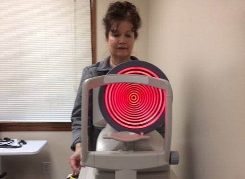
Corneal Topography
Corneal topography is a computer assisted diagnostic tool that creates a three-dimensional map of the surface curvature of the cornea. The three-dimensional map is a valuable aid to the examining optometrist and can assist in the diagnosis and treatment of a number of conditions; in planning cataract surgery and intraocular lens (IOL) implantation (plano or toric IOLs); in planning refractive surgery such as LASIK, and evaluating its results; or in assessing the fit of contact lenses.
The procedure is safe, non-invasive and takes only seconds to complete thus allowing your doctor to make educated and detailed decisions about your eye care. Corneal topographer technology is also extremely useful for laser vision correction because of the curvature reshaping of your corneas for clearer vision.
Optomap Ultra-Widefield Retinal Image
A non-invasive, low-powered scanning laser, Optomap digitally scans the retina allowing your doctor to identify potential retinal problems such as macular degeneration, glaucoma, retinal holes or detachments.
The optomap ultra-widefield retinal image is a unique technology that captures more than 80% of your retina in one panoramic image while traditional imaging methods typically only show 15% of your retina at one time.
The benefits of having an optomap ultra-widefield retinal image taken are:
- Optomap facilitates early protection from vision impairment or blindness
- Early detection of life-threatening diseases like cancer, stroke, and cardiovascular disease
The unique optomap ultra-widefield view helps your eye care practitioner detect early signs of retinal disease more effectively and efficiently than with traditional eye exams
Heidelberg Retina Tomography (HRT)
The key to controlling glaucoma is catching it early. Using the Heidelberg Retina Tomography technology we are able to diagnose glaucoma years before symptoms become apparent to the patient. HRT is a system that combines a laser-scanning camera with specialized software allowing our doctors to accurately evaluate the optic nerve. In only moments, the HRT gives measurements of the size, depth and shape of the optic nerve and can easily detect small changes that occur over time. It is a painless, non-invasive test that rarely calls for eye dilation. This advanced technology is a quick and accurate way to follow your progress from exam to exam.
Optical Coherence Tomographer (OCT)
Our OCT is a safe, painless and non-invasive way to obtain detailed images of the retina or optic nerve. OCT helps us better manage glaucoma and diseases of the retina because this technology allows the eye doctor to see the deep tissue layers in the eye. Similar to ultrasound, this diagnostic technique employs light rather than sound waves to achieve higher resolution pictures of the structural layers of the back of the eye. These high-definition images are the only way that they can actually see beneath the surface to the nerve fiber layers where damage occurs. Up until now, eye doctors had to use other tests to indicate damage in this critical area of sight. Common eye diseases such macular degeneration, diabetic retinopathy, and glaucoma are detected early by the OCT when the diseases can be more effectively treated.
Corneal Pachymetry
The corneal pachymeter uses sophisticated ultrasound technologies to gently and accurately measure the eye’s corneal thickness in micrometers. Correctly measuring the corneal thickness is crucial because it can mask correct readings of eye pressure, potentially causing a misdiagnosis. We also use this instrument to aid in the diagnosis and treatment of corneal diseases as well as for prospective laser vision correction candidates.